Whole Leaf Aloe Vera And
|
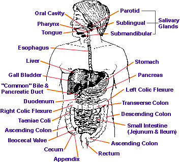
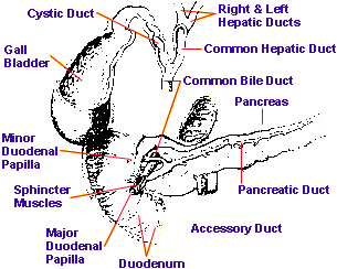
However, too much acid can be a serious disadvantage also, as we shall see below. This phase may characteristically last for about 2 hours before the stomach starts to empty, but is very variable. In particular the time of residence in the stomach is lengthened by a high fat content in the meal, which may delay emptying for quite a long time. When the stomach empties, its contents are passed on into the duodenum, which is the first part of the small intestine. Here the very acid, partly digested, fluid material, now called “chyle,” meets the pancreatic juice and the bile, which are both secreted into the duodenum, respectively from the exocrine pancreas and from the liver and gall bladder, (digest fats), trypsin, chymotrypsin and carboxypeptidase (to continue the digestion of proteins) and pancreatic amylase (to continue the digestion of starch). The pancreatic juice therefore amounts to a quite formidable battery of enzymes able to break down all the main bulk nutrients. The bile contains many wastes and toxins, for it is one of the functions of the liver to clear the blood of toxins and excrete them into the bile for passing out of the body. However, it also contains the bile salts, taurocholic and glycocholic acids, which are potent fat emulsifiers. These play an important part in fat digestion by breaking down the larger fat droplets into smaller ones. The duodenum is in many ways the hub of the digestive process, where numerous key steps are concentrated. It is extremely important that the control of pH within the duodenum should be correct. The pancreatic enzymes have their working optimum on the alkaline side of neutrality, so they cannot work properly if the combined effect of the slightly alkaline bile and the pancreatic juice should fail to neutralize the strong acid of the chyle. Under these conditions, the chyle will remain acid and the intestinal phase of digestion cannot get properly underway. The situation will also expose the relatively delicate tissues of the duodenum to un-neutralized acid from the stomach and may encourage ulceration of the duodenum. Digestion and absorption normally proceed, with fats being emulsified and partly broken down by pancreatic lipase, to be absorbed further down the small intestine, partly as fatty acids and glycerol and partly as tiny fat droplets which go into the blood as “chylomicrons.” Proteins are attacked extensively by the pancreatic proteases as intestinal digestion proceeds, and are joined by other enzymes which break down smaller peptides, some of these enzymes being produced in the intestinal juice itself otherwise known as the “succus entericus.” Eventually, proteins are reduced to free amino acids and absorbed. Starches are reduced mainly to maltose, a disaccharide which has then to be broken down to glucose by the action of the maltase enzyme in the succus entericus. Common sugar or sucrose, is split by sucrase from the succus entericus. As the food passes to the jejunum, (the mid part of the small intestine) and the ileum (the final part of the small intestine), these various digestive and absorptive processes begin to approach completion.
In the large intestine, or colon, much water is reabsorbed, which is a very important function. With this, the colon also reabsorbs many important mineral salts. This reabsorption of mineral salts is significant because, although much absorption of minerals also occurs in the small intestine, this is never complete. This is more than just the absorption of dietary minerals. The digestive juices are mineral rich. If any significant proportion of the mineral reserves that are “invested” in the digestive process failed to be reabsorbed, that would represent a serious loss to the body. This is prevented by having a colon which is competent at mineral absorption. Under the best conditions, some small proportion of the total starch intake will remain by the time the food residues reach the colon. This will then provide an energy source for the Lactobacillus acidophilus and other desirable acid-forming bacteria. These, if they are well established there, will inhibit the growth of undesirable putrefactive bacteria and even pathogens, and are known to have some anti-tumour properties. They will also manufacture significant amounts of vitamins which supplement dietary sources of vitamins. High protein content should never be allowed to reach the colon, since it will lead to the production of alkali rather than mild acid. This will favour the undesirable putrefactive bacteria, pathogens and Candida albicans, and, through the decarboxylation of amino acids, will produce quantities of toxic amines which become absorbed and intoxicate the body and all the organs within it. Disturbances Of DigestionSo, digestive disturbance may begin from either too much acid or too little acid and pepsin in the stomach. If the stomach phase of digestion is less effective than it should be, then protein may well pass down into the lower bowel to undergo putrefaction and an overwhelming production of toxins. That is doubly likely if the pancreas is also sluggish or incompetent in the production of an enzymatically active pancreatic juice. The condition of both stomach and pancreas can be read diagnostically in the iris of the eye.When putrefaction sets in, the intestines themselves become compromised and are often ineffective in their normal functions. They are liable to become pocketed, bulged, and affected by diverticuli. Their ability to carry out peristalsis (the muscular movements which advance the food residues along the intestine), becomes sluggish, the tissues of the intestinal wall become toxic, weakened and vulnerable to infection and ulceration. These effects are obviously going to be noticed eventually in terms of bowel diseases of one kind or another. High up in the intestine there is danger of ulceration wherever a substantial excess of un-neutralized acid prevails, over and above that which is required in any part of the gastrointestinal system. There is obviously a strong correlation between over-acidity and the occurrence of either gastric or duodenal ulcers - even though some other factors may have to be present also to cause breakdown of the normal protection of the stomach or duodenal wall. In the small intestine, conditions of inflammation and/or abnormal levels of secretion may well occur if the pH of the contents are wrong or if the small intestinal tissues are not being properly nourished through errors of the digestive process higher up in the tract, especially errors of function in the stomach, liver or pancreas. What has been described above is a maze of possible symptoms that may be cross-connected in diverse ways. Whilst some improvements may sometimes be gained by a piecemeal and symptomatic approach, a wholistic approach to the overall working of the digestive system, as has already been stated, is far more likely to provide a truly effective and lasting solution. To gain insight into how Aloe affects the working of the digestive system as a whole, it is necessary to consider at some length the work of Dr. Jeffrey Bland as reported in his paper “Effect of Orally Consumed Aloe vera juice on Gastrointestinal Function in Normal Humans.” Dr Bland wrote this paper from the Linus Pauling Institute of Science & Medicine at Palo Alto, California. It was published in Preventive Medicine in the Issue of March / April 1985. In the tests reported by Bland, the dose of unconcentrated Aloe vera juice was 6 ounces per day (i.e. about 170ml), divided into 3 aliquots of 2 ounces (59ml). The duration of the test was only 7 days and no special measures were taken with regard to diet during the test period. Several parameters were measured which, taken together, were regarded as providing as a good and reliable index of the functioning of the gastrointestinal system. These were (1) a stool culture to indicate the distribution of bacterial types (2) levels of indican in the urine as an indication of the putrefactive capability of the intestinal flora and hence of the flora’s capacity to manufacture toxic amines from intestinal amino acids (3) stool density (4) bowel transit time and (5) gastric pH. The results indicated about a 40% reduction in the indican levels. This was taken to indicate that either bowel putrefactive activity was reduced, or else the digestion and assimilation of dietary protein higher up the tract was improved, or possibly both. Indican is derived from the amino acid tryptophane, but it was being used is a likely indicator of overall amino acid decarboxylating activity, and therefore of toxic amine production generally. The markedly diminished indican levels in the urine were taken, quite correctly, I think, to represent a considerable improvement in overall gastrointestinal function. It is a finding which carries with it implications for gastric function, pancreatic function, better bowel flora composition and, correlated to that, bowel contents pH and lower putrefective activity. The stool cultures indicated an improved composition of the bacterial flora of the gut following the Aloe vera test. It is interesting that this improvement was attained without the use of bowel flora products containing supplements of live bacteria. Clearly, the Aloe vera itself was creating conditions within which a better spectrum of bacteria could survive and grow. The advantages of this are well known to nutritionists, and are clearly linked to lower putrefactive activity as outlined above. One especially interesting finding was that the yeast count in the stool cultures diminished markedly. The specific gravity of the stools was reduced on average by 0.37 units. This was interpreted as an important shift towards a more ideal value. It was taken to indicate a better water-holding capacity of the stools and a faster transit time through the gastrointestinal system. It was reported that no-one suffered from diarrhea or loose stools during the test. Clearly, the Aloe vera was not acting as a laxative at all. The better bowel transit time was interpreted as an improvement of muscular tone throughout the gastrointestinal system. The study clearly established that Aloe vera exerted a marked effect upon gastrointestinal pH. Whilst this was profoundly interesting, it was the least satisfactory part of the study because the pH changes in different sections of the gastrointestinal tract were not separately reported and differentiated. However, Bland’s tabulated results suggest that a reduction in average gastric acidity was the most pronounced finding, being a reduction by 1.88 pH units. In accord with explanations I have given above, a reduction in stomach acidity will only be of benefit to people who originally had hyperacidity. It is noticeable in Bland’s results that two individuals with a starting gastric acidity of less than pH 2 (i.e., very acid), showed a pH change of 2.55 units whilst those with a relatively non-acid pH of above 4 only showed an average change of 0.45 units. It appears, therefore, that people who experienced major change of gastric pH were the people who really needed on account of previous hyperacidity. Although the subjects for this study were “normal humans,” the explanations given earlier in this fact sheet make it clear just why these people would have been closer to possible gastrointestinal upset than the others and also make it clear that the observed reduction in gastric pH would have been beneficial. It also becomes clear that here also is one reason why, in abnormal human subjects, conditions of gastric and duodenal ulceration would be much relieved by Aloe vera juice. It now seems clear that the combined effect of all these various parameters of function should be taken into account when assessing the effect of Aloe upon gastrointestinal function. Thinking piecemeal, symptomatically and non- wholistically is just not good enough to generate the level of understanding required. No other studies appear to vie with the Bland study for detailed monitoring and whole-system investigation. More such studies are obviously needed in which Aloe vera is used for rather longer and in which people with named digestive abnormalities are included in the study. Conditions such as colitis, diverticulitis, ulcerative colitis, Crohn’s disease and irritable bowel syndrome (IBS) specifically need to be investigated. From what is known of the nature of these complaints and what is known of the actions of Aloe vera, there is every reason to expect such trials to be positive. A great many Alternative Practitioners, working with their individual patients, are already informally reporting success with these named complaints. There is a scientific study from the Ukraine which concluded very positively that “In cases of functional disorders of the small intestine the process of juice secretion and enzymatic activity, Aloe extract may be recommended for stimulating the secretory function of the small intestine.” This suggests that a small intestinal condition such as Crohn’s disease is likely to be helped. The fact that in this case the Aloe was injected may not, of course, be essential to its efficacy. Peptic UlcerSome Japanese work concerns peptic ulcer, as does the work of Blitz and colleagues in Florida (1963). In the latter study 12 patients with peptic ulcer were selected and Aloe vera gel was the sole source of treatment. It is notable that the gel was used by Blitz because in the Japanese work some components of the exudate fraction of the leaf (which is absent from gel) were recognised as being important. The twelve patients were “diagnosed clinically as having peptic ulcer, and having unmistakable roentgenographic evidence of duodenal cap lesions.” The results of the Blitz work are summarized as “All of these patients had recovered completely by the end of 1961, so that at this writing at least 1 year has elapsed since the last treatment.” Also “Clinically, Aloe vera gel has dissipated all symptoms”; and “Aloe vera gel provided complete recovery.” It is, indeed, tantalizing when one has only a small quantity of good information on such an important subject. The chances are that the misery of thousands of peptic ulcer sufferers could be removed through Aloe vera, but no one has proved it on a large enough scale, or to the satisfaction of the medical profession. The lucky members of the public are the ones who know about it.Another study in 1978 is significant insofar as it identifies in several papers that two factors in Aloe which diminish stomach secretion are, aloenin and Aloe-ulcin. They obtained these from Aloe Arborescens. Aloenin is one of the individual components of the exudate fraction of the leaf. It is a phenolic compound of the type called a “quinonoid phenylpyrone.” The fact that aloenin has this property means that it would have an action not unlike that of a drug such as cimetedine, marketed as Tagamet, which has a huge usage as a chemical drug for the treatment of peptic ulcer by suppression of stomach secretion. It is to be hoped that the action of substances from the gel or whole leaf extract upon peptic ulcer will be found to be by a less crude and less suppressive mechanism, which, hopefully might have something to do with correcting the underlying causes of peptic ulcer. Nonetheless, the Japanese findings show that, a named component of the exudate fraction (aloenin) seems to have a synergistic effect (i.e. a mutually enhancing effect) with the action of the other leaf components. As for Aloe-ulcin, the Japanese identified it with magnesium lactate. It is, frankly, hard to become convinced by that part of the evidence, because there is so little magnesium in Aloe: it takes much more to have known physiological effects. Therefore, this author does not draw any firm conclusions about Aloe-ulcin, but this need not affect, in any way, the overall conclusions in relation to peptic ulcer. The clinical evidence, both from the work of Blitz and from the Japanese work, is clear, in spite of their small numbers of patients. The effectiveness of Aloe Vera for peptic ulcer seems established, even if some component of the exudate, such as aloenin, might ideally be added for maximum effect. There is, in my view, quite enough evidence to support the use of Aloe vera Whole Leaf Extract as a component of treatment for every peptic ulcer case encountered. This completes the case in favor of using Aloe vera Whole Leaf Extract for maintaining, improving and healing the digestive tract. |
Whole Leaf Aloe Vera Human Digestive System
Keywords for this page: Psoriasis, Psoriasis Treatment Protocol, Aloe Vera, Whole Leaf, wholeleaf, Cold Processed, Aloe Barbadensis plant, Acemannan, Whole Leaf Aloe Vera Beverage, Cold processed Whole leaf, Mucopolysaccharides, MPS, scaly, shiny lesions of skin
Whole Leaf Aloe Vera - Home Whole Leaf Aloe Vera Products Aloe Vera Information

