Aloe And Other Topical Antibacterial Agents
In Wound Healing

 
By John P. Heggers, Ph.D. & Wendell Winters, Ph.D.


Unlike any other wound, the burn is a non-uniform injury in which some tissues are partially or completely damaged, while other tissues suffer minimal damage. The latter will heal without any therapeutic treatment, while the former will become permanently damaged, creating a granulating wound if not appropriately treated.

Infection also plays a major role in the conversion of this wound.1 Many of the topical agents used are to control burn wound infections. However, there are other products that have multivaried effects on the burn wound. Some of the major properties attributed to Aloe vera include its ability to:

  1. penetrate tissue
  2. anesthetize the tissue
  3. allay bacterial, fungal & viral growth
  4. act as an anti-inflammatory
  5. dilate capillaries & enhance blood flow2

Heggers and his co-workers3 showed that topical application of anti-eicosanoids, more specifically anti-thromboxane agents, could reverse progressive tissue necrosis in the partially damaged tissue. Topical application of an Aloe compound resulted in healing patterns comparable to the anti-thromboxane agents. Robson and his colleagues2 also showed that such an Aloe compound had anti-bacterial properties as well.

Therefore, topical application of anti-microbials and other chemo-therapeutic agents is essential in order to restore the normal healing process and prevent infection. Halsted has been quoted as saying, “A wound which has been irrigated with solutions of carbolic acid, corrosive sublimate, or other disinfectant labors under the disadvantage of a more less extensive area of superficial necrosis”3. McCauley and his colleagues4 have show that both silver sulfadiazine and Sulfamylon® are toxic to fibroblasts in tissue culture at concentrations of 0.005% and 0.1%, respectively. Leitch, et al5 recently presented data that silver sulfadiazine, Sulfamylon® and silver sulfadiazine with chlorohexadine significantly retarded wound healing in the acute wound model.

Since application of topical chemotherapeutic agents is essential in the prevention of infection and enhancement of wound healing, we examined Aloe’s role in accelerating wound healing or reversing the wound retardant effect of silver sulfadiazine as well as the influence of Bactroban® and clindamycin on the healing process.

In order to be assured that Aloe contained active components which are essential in the healing process we employed a Polyarcylamide gel electrophoresis (PAGE) and cell growth assays to determine the presence of active components. Acrylamide-bis acrylamide 37.5:1 (Fisher Biotech Houston), tris-HCI (Sigma St. Louis), sodium lauryl sulfate (SDS), N,N,N’,N’-tetramethylenediamine (TEMED), ammonium persulfate (Bio-Rad (Richmond), rainbow weight marker (Amersham Arlington Heights were purchased from the respective vendors). Electrophoresis was performed using a 12.5% separating gel and a 4% stacking gel run in a Bio-Rad Protean II Xi vertical electrophoresis cell system at 3OmA constant current6. The gel was stained by silver stain kit method, and the kit was purchased from Bio-Rad.7

Cells were grown in Dulbecco’s Minimal Medium (DMEM) supplemented with 10% heat-inactivated horse serum, 5% fetal calf serum, 50 units penicillin, 0.05% mg/ml streptomycin 1mM L-glutamine and 1mM sodium pyruvate. The rat adrenal cultured cells were prepared following incubation at 37o C in 5% CO2. Cell concentration and viability were determined by hemacytometer counts and dye exclusion with 0.04% trypan blue.

Cells at 5x10⁴ cell/well were plated into 96-well flat-bottom plates and maintained 24 hours at standard conditions in adherence studies. The media was removed before the addition of the Aloe-DMEM mixture. After 72 hours of incubation, 10ul of MTT (3-[4,5-Dimeth-ylthiazol-2-yl]-2,5-diphenyl-tetrazolium bromide) solution (5mg/ml) was added to each well. The formazan crystals, formed only in viable cells after four hours at 37o C, were dissolved by addition of 100ul of acid-isopropanol solution. The plates were read by a Micro ELISA reader (MR 580 Dynatec) at 570nm (630nm reference wavelength, calibration setting of 1.99).

The acute model was used as previously described by Heggers, et al.8 Appropriately anesthetized Sprague Dawley rats, two proximal and two distal, received four 1.5 cm2 dorsal defects through the skin and panniculus carnosus. This study was conducted in compliance with UTMB’s Animal Care and Use Committee under ACUC protocol #92-05-026. (Fig. 1)

Figure 1

Figure 1 Schematic representation of the acute wound healing model.

The skin defects were treated three times a day for 14 days with Aloe vera gel (n=10), 2% mupirocin ointment (n=10), 1% clindamycin cream (n=10), 1% silver sulfadiazine alone (n=10), 1% silver sulfadiazine cream + Aloe (n=10). An untreated group served as control (n=10). Wound closure rate was assessed by serial planimetry. Following healing, the breaking strength of each resultant scar was determined using an Instron tensiometer model #4201 (Instron Corp, Canton, MA). Wound half-lives and overall healing rates were calculated by regressing the log of the areas of all wounds over time.

Figure 2
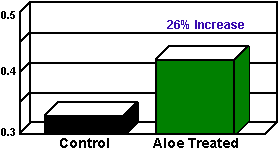
Figure 2
Tissue culture response to Aloe 1:1 gel
compared to control (untreated).

Results

The SDS PAGE analysis revealed a high molecular weight polypeptide in Aloe vera 1:1 gel #5.

The rat adrenal cultured cells in the presence of Aloe vera gel #5 showed a 26% increase in growth activity when compared to the control (Fig. 2). Therefore, we utilized the Aloe vera gel #5 for our in vivo assay.

Acute Wound Healing

Topical application of each therapeutic agent had a profound effect on the healing process. Overall healing rates of all the treated groups were significantly different as compared to the control group (p<0.05). The Aloe group had the shortest half-life, and healed faster than the control group (Table I). All the other treated groups had longer half-lives compared to the control group. While silver sulfadiazine with Aloe significantly increased the breaking strength (2.000 + 0.504) of the healed wound, Aloe alone was slightly stronger than the control silver sulfadiazine.
Table I
Fractional Area & Healing Rates of Wounds Treated With Topical Antibacterials
Group
Days
(n)
Fraction Of Initial Wound
Area Throughout + SD
Overall Healing
Rate (Slope +SD)
1/2 Life
1. Control
480
0.289 + 0.385
0.1477 (0.0027)
6.38
2. Aloe
360
0.279 + 0.364
0.1657 (0.0027)
6.14*
3. SSD
480
0.368 + 0.420
0.1800(0.0050)
8.56
4. SSD + Aloe
480
0.277 + 0.392
0.1339 (0.0030)
6.94
5. Bactroban®
480
0.332 + 0.414
0.1300 (0.0026)
8.74
6. Clindamycin
324
0.396 + 0.482
0.1711 (0.0037)
8.30
*All half-life days are significant (p = <0.05)
Table II
Breaking Strength of Healed Wounds
Group
Breaking Strength
(KG) + S
(n)
1. Control 1.461 + 0.421 30
2. Aloe 1.640 + 0.533 29
3. SSD 1.521 + 0.432 28
4. SSD + Aloe 2.000 + 0.504* 28
5. Bactroban® 1.845 + 0.421 24
6. Clindamycin 1.621 + 0.404 15
*SSD + Aloe breaking strength is significant (p = <0.05)


Topical Aloe significantly enhances the rate of wound healing, and, when combined with silver sulfadiazine, it apparently reverses the wound retardant effect of silver sulfadiazine. Clindamycin and mupirocin significantly delayed wound closure as did silver sulfadiazine, while the breaking strength for the three topical agents appears stronger or comparable to the control. (Table II)


Conclusions

Topical application of a variety of cytokines to open wounds has revolutionized the process of wound healing. Hayward, et al9 provided evidence that the basic Fibroblast Growth Factor (bFGF) reverses bacterial retardation of wound contraction in a chronic granulating wound.

Carney and his co-workers10 showed that exogenous delivery of synthetic Thrombin Receptor-activating peptides enhanced the healing process and neovascularization of an incisional wound. In a clinical trial Bishop, et al11 evaluated two potential wound healing agents in a blinded trial for the treatment of venous status ulcers. Contrary to previous in vitro and in vivo studies by McCauley, et al4 and Leitch, et al5 the Bishop study showed that silver sulfadiazine was significantly more therapeutic in healing the venous status ulcer when compared to a biologically active tripeptide copper complex or a placebo. These results suggest that a silver sulfadiazine cream may facilitate healing in wounds that heal by epitheliazation. Robson and co-workers,12, 13 in two separate publications, reported on the efficacy and safety of platelet-derived growth factor B-B and bFGF in chronic pressure sore ulcers.

Our previous studies have provided evidence that Aloe vera may contain a growth factor like substance.2, 3, 4 Recently, Winters14 presented data regarding a growth stimulating as well as a growth suppressant substance in Aloe gel. PAGE analysis revealed the presence of a polypeptide species possibly responsible for these activities. By immunoblot technique, the Aloe substances were found to contain Na+/K+ATPase and Con A activities. These mitogenic lectin-like substances in Aloe have been previously described.

Therefore, with this foundation of knowledge regarding the exogenous administration of cytokines and Aloe substances in the process of wound healing, we closely examined Aloe vera gel 1:1 (#5) to provide further evidence of its wound-healing potential compared to other chemotherapeutic agents.

McCauley, et al4 showed that silver sulfadiazine in tissue culture was toxic to fibroblasts and keratinocytes, and Leitch and his co-workers5 showed that it retarded wound healing in vivo. We wondered if Aloe would respond as bFGF did in reversing the retardation of wound healing9 and if the treated, healed wound was stronger or weaker than the control wound.

The Aloe-alone treated wounds healed faster and had a half-life of 6.14 days which was significantly (p<0.05) shorter when compared to the other groups (Table 2). The silver sulfadiazine and Aloe group, while it healed significantly faster (p = <0.05) than the Bactroban® silver sulfadiazine, clindamycin groups, had a half-life of 6.94 which was slightly longer than the control wound (half-life 6.38).

The breaking strength of the Aloe-treated wound (1.640) was significantly less (p<0.05) than the silver sulfadiazine + Aloe group (2.000) (Table II). The control breaking strength was 1.461, less than the Aloe-treated wounds, while the silver sulfadiazine + Aloe treated wounds were significantly (p<0.05) greater than all other groups. The Bactroban® -clindamycin- and silver sulfadiazine-treated wounds were apparently stronger than the controls, but the healing time was significantly (p<0.05) retarded. This study further substantiates the fact that Aloe contains a growth promoting factor that enhances the healing process and the breaking strength of these healed wounds. Aloe can also reverse the wound healing retardant effect of silver sulfadiazine, a topical antimicrobial used to treat and control burn wound sepsis.

References

Whole Leaf Aloe Vera - Aloe And Other Topical Antibacterial Agents In Wound Healing


Whole Leaf Aloe Vera - Home
Whole Leaf Aloe Vera Products
Aloe Vera Information
Aloctin A, An Active Substance Of Aloe Arborescens Miller As An Immunomodulator
Aloe Vera - Whole Leaf Aloe Vera - Wound Care Protocol
Aloe Vera Juice - Effect Of Orally Consumed Aloe Vera Juice
Aloe Vera Leaf Handling And Constituent Variability
Aloe Vera, The Whole Leaf Advantage
Fundamental Healing Properties Of Aloe Vera Mucopolysaccharides
Immune Enhancing Effects Of Whole Leaf Aloe Vera
Internal Uses Of Aloe Vera
Internal Uses Of Aloe Vera - References
Psoriasis | Psoriasis Treatment Aloe Vera
The Healing Properties Of Whole Leaf Aloe Vera
The Rediscovery Of Aloe Vera: Cold Processed Whole Leaf Aloe Vera
Whole Leaf Aloe Vera - Aloe And Other Topical Antibacterial Agents In Wound Healing
Whole Leaf Aloe Vera - Aloe And Other Topical Antibacterial Agents In Wound Healing - References
Whole Leaf Aloe Vera - Aloe In Alternative Medicine Practice
Whole Leaf Aloe Vera - Aloe Medicinal Substances
Whole Leaf Aloe Vera - Aloe Vera Gel In Peptic Ulcer Therapy
Whole Leaf Aloe Vera - Aloe Vera Gel In Peptic Ulcer Therapy - References
Whole Leaf Aloe Vera - Aloe's Effectiveness As An Anti-Inflammatory Agent
Whole Leaf Aloe Vera - Aloes In The Treatment Of Burns And Scalds
Whole Leaf Aloe Vera - An Orchestra of Active Compounds
Whole Leaf Aloe Vera - Ancient Herb In New Form Delivers Proven Effects
Whole Leaf Aloe Vera - Antiarthritic Activity Of Anthraquinones Found In Aloe Vera For Podiatric Medicine
Whole Leaf Aloe Vera - Anti-Inflammatory Activity In Diabetes
Whole Leaf Aloe Vera - Anti-Inflammatory Activity In Diabetes - References
Whole Leaf Aloe Vera - Biological Activity Of Aloe Vera
Whole Leaf Aloe Vera - Polysaccharides and Wound Healing
Whole Leaf Aloe Vera - Prevention Of Atheromatous Heart Disease
Whole Leaf Aloe Vera - The Cancer Solution
Whole Leaf Aloe Vera - A Holistic Protocol For The Immune System
Whole Leaf Aloe Vera - The External Use Of Aloes
Whole Leaf Aloe Vera - Use In Wound Healing And Disease Control In Oral Conditions
Whole Leaf Aloe Vera - Wound Healing, Oral And Topical Activity Of Aloe Vera
Whole Leaf Aloe Vera - Wound Healing, Oral And Topical Activity Of Aloe Vera - References
Whole Leaf Aloe Vera and Drug-Resistant Bacteria, Viruses & Fungi
Whole Leaf Aloe Vera And The Heart
Whole Leaf Aloe Vera And The Heart - References
Whole Leaf Aloe Vera And The Human Immune System
Whole Leaf Aloe Vera And The Skin
Whole Leaf Aloe Vera Beverage - Buy here on-line
Whole Leaf Aloe Vera Human Digestive System
Whole Leaf Aloe Vera In Dentistry
Whole Leaf Aloe Vera Tumor Inhibitors Aloe Emodin
Whole-Leaf Aloe Vera - Digestion and The Immune System
Whole-Leaf Aloe Vera, Almost A Panacea - References
Whole-Leaf Aloe Vera, Almost A Panacea
Site Map